Marek Gowkielewicz, Aleksandra Lipka, Wojciech Zdanowski, Tomasz Waśniewski, Marta Majewska, Carsten Carlberg
Anti-Müllerian hormone: biology and role in endocrinology and cancers
Front. Endocrinol., 16 September 2024, Sec. Cancer Endocrinology, Volume 15 - 2024

Anti-Müllerian hormone (AMH) is a peptide belonging to the transforming growth factor beta superfamily and acts exclusively through its receptor type 2 (AMHR2). From the 8th week of pregnancy, AMH is produced by Sertoli cells, and from the 23rd week of gestation, it is produced by granulosa cells of the ovary. AMH plays a critical role in regulating gonadotropin secretion, ovarian tissue responsiveness to pituitary hormones, and the pathogenesis of polycystic ovarian syndrome. It inhibits the transition from primordial to primary follicles and is considered the best marker of ovarian reserve. Therefore, measuring AMH concentration of the hormone is valuable in managing assisted reproductive technologies. AMH was initially discovered through its role in the degeneration of Müllerian ducts in male fetuses. However, due to its ability to inhibit the cell cycle and induce apoptosis, it has also garnered interest in oncology. For example, antibodies targeting AMHR2 are being investigated for their potential in diagnosing and treating various cancers. Additionally, AMH is present in motor neurons and functions as a protective and growth factor. Consequently, it is involved in learning and memory processes and may support the treatment of Alzheimer’s disease. This review aims to provide a comprehensive overview of the biology of AMH and its role in both endocrinology and oncology.

View full text
​Ranjini Ghosh Dastidar, Julia Jaroslawska, Marjo Malinen, Tomi‑Pekka Tuomainen, Jyrki K. Virtanen, Igor Bendik & Carsten Carlberg
In vivo vitamin D targets reveal the upregulation of focal adhesion‑related genes in primary immune cells of healthy individuals
Scientific Reports volume 14, Article number: 17552 (2024), 30 July 2024

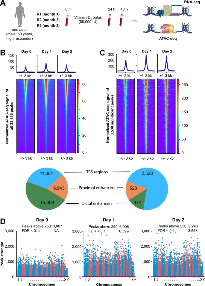
Vitamin D modulates innate and adaptive immunity, the molecular mechanisms of which we aim to understand under human in vivo conditions. Therefore, we designed the study VitDHiD (NCT03537027) as a human investigation, in which 25 healthy individuals were supplemented with a single vitamin D3 bolus (80,000 IU). Transcriptome-wide differential gene expression analysis of peripheral blood mononuclear cells (PBMCs), which were isolated directly before and 24 h after supplementation, identified 452 genes significantly (FDR < 0.05) responding to vitamin D. In vitro studies using PBMCs from the same individuals confirmed 138 of these genes as targets of 1α,25-dihydroxyvitamin D3. A subset of the 91 most regulated in vivo vitamin D target genes indicated focal adhesion as the major pathway being upregulated by vitamin D3 supplementation of healthy individuals. Differences in the individual-specific responsiveness of in vivo vitamin D target genes in relation to the increase of the person’s vitamin D status allowed a segregation of the VitDHiD participants into 9 high, 12 mid and 4 low responders. The expression profile of nearly 600 genes elucidate the difference between high and low vitamin D responders, the most prominent of which is the HLA-C (major histocompatibility complex, class I, C) gene.

View full text
​Julia Jarosławska, Ranjini Ghosh Dastidar, Carsten Carlberg
In vivo vitamin D target genes interconnect key signaling pathways of innate immunity
PLoS ONE July 2024 19(7): e0306426

The vitamin D3 metabolite 1,25-dihydroxyvitamin D3 (1,25(OH)2D3), its nuclear receptor VDR (vitamin D receptor) and hundreds of their target genes are not only key regulators of calcium homeostasis, but also important modulators of the immune system. Innate immune cells like monocytes use VDR for efficient differentiation and are very responsive to vitamin D. So far, most information on the gene regulatory function of vitamin D and its physiological impact had been obtained from in vitro studies using supraphysiological doses of 1,25(OH)2D3. Therefore, medical experiments like the study VitDHiD (NCT03537027), where 25 healthy individuals were supplemented once with a vitamin D3 bolus (80,000 IU), provide important insight into the response to vitamin D under in vivo conditions. In this study, we inspected 452 in vivo vitamin D target genes from peripheral blood mononuclear cells (PBMCs) detected in VitDHiD and found 61 of them involved in eight major KEGG (Kyoto Encyclopedia of Genes and Genomes) pathways of innate immunity. Under in vivo conditions in healthy individuals vitamin D either silences five pathways of innate immunity, stabilizes two and increases one, so that acute inflammation is suppressed and the release of cytokines is kept under control. A ranking of the 61 target genes by inducibility, basal expression and multiple involvements in the pathways highlighted the genes NFKBIA (NFκB inhibitor alpha), NFKBIZFOSL2 (FOS like 2, AP1 transcription factor subunit), JDP2 (Jun dimerization protein 2), PIK3R1 (phosphoinositide-3-kinase regulatory subunit 1), CLEC7A (C-type lectin domain containing 7A), DUSP6 (dual specificity phosphatase 6), NCF2 (neutrophil cytosolic factor 2), PLCB1 (phospholipase C beta 1), PLCG2 and TNFAIP3 (TNF alpha induced protein 3). In conclusion, vitamin D’s in vivo effect on innate immunity in healthy adults is mediated by the interconnection of the pathways of neutrophil extracellular trap formation, Toll-like receptor, chemokine and phagosome signaling, NOD-like receptor, C-type lectin receptor, apoptosis and interleukin 17 through a limited set of proteins encoded by key target genes.

View full text
Eunike Velleuer, Carsten Carlberg
A Nutrigenomic View on the Premature-Aging Disease Fanconi Anemia
Nutrients 2024, 16(14), 2271

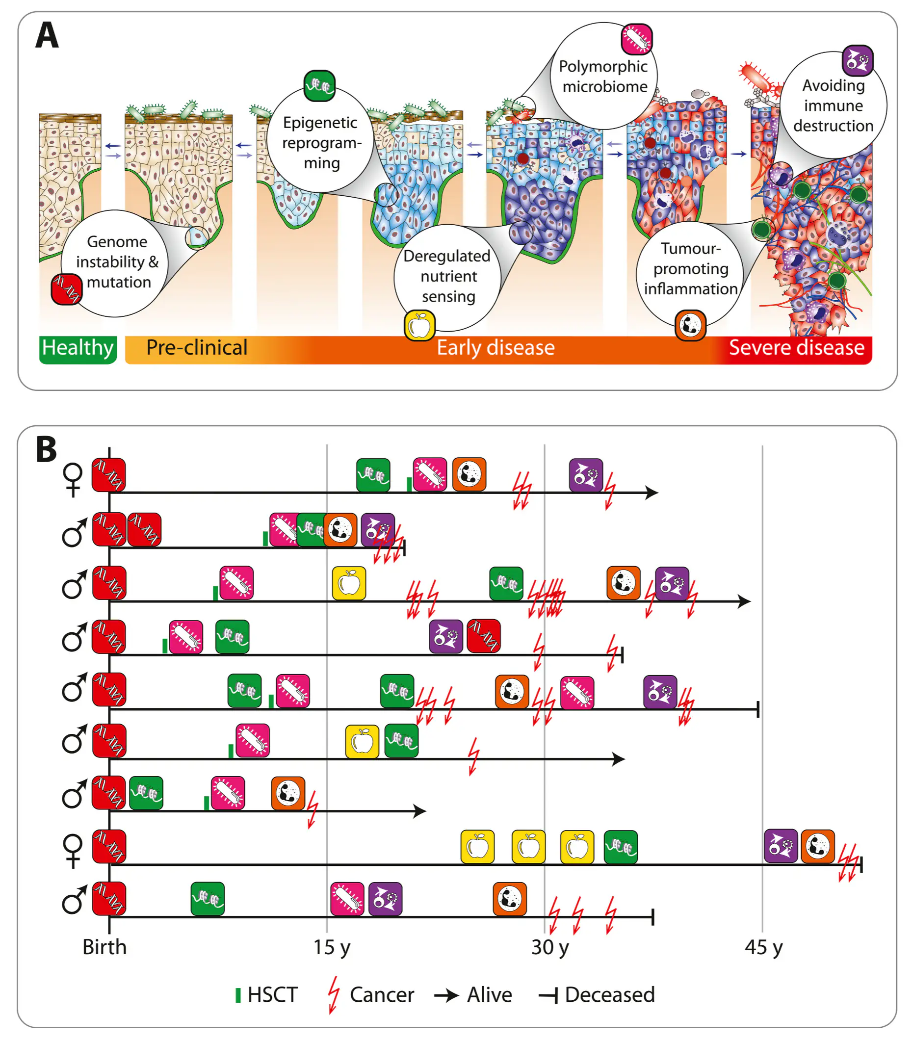
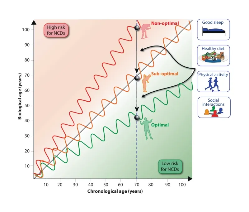
Fanconi anemia, a rare disorder with an incidence of 1 in 300,000, is caused by mutations in FANC genes, which affect the repair of DNA interstrand crosslinks. The disease is characterized by congenital malformations, bone marrow failure within the first decade of life, and recurrent squamous cell carcinomas of the oral cavity, esophagus, and anogenital regions starting around age 20. In this review, we propose that Fanconi anemia should be considered a premature-aging syndrome. Interestingly, the onset and severity of the life-limiting clinical features of Fanconi anemia can be influenced by lifestyle choices, such as a healthy diet and physical activity. These factors shape the epigenetic status of at-risk cell types and enhance the competence of the immune system through nutritional signaling. Fanconi anemia may serve as a model for understanding the aging process in the general population, addressing research gaps in its clinical presentation and suggesting prevention strategies. Additionally, we will discuss how the balance of genetic and environmental risk factors—affecting both cancer onset and the speed of aging—is interlinked with signal transduction by dietary molecules. The underlying nutrigenomic principles will offer guidance for healthy aging in individuals with Fanconi anemia as well as for the general population.

View full text